| Info
Sheets |
| | | | | | | | | | | | | | | | | | | | | | | | |
 | Out-
side |
| | | | |
|
| | | | |
Result : Searchterm 'Perfusion Imaging' found in 1 term [ ] and 15 definitions [ ] and 15 definitions [ ], (+ 8 Boolean[ ], (+ 8 Boolean[ ] results ] results
| | previous 16 - 20 (of 24) nextResult Pages : [1] [2 3 4] [5] |  | |  | Searchterm 'Perfusion Imaging' was also found in the following services: | | | | |
| |  |
| |
|
Device Information and Specification
CLINICAL APPLICATION
Whole body
CONFIGURATION
Mobile compact
Whole body, intra-operative head, neck volume, atlas head//neck vascular quadrature phased array, spine quadrature, C/T/L spine phased array, small joint, large joint, TMJ bilateral, shoulder phased array, extremity quadrature volume, wrist, hand quadrature, general purpose flexible, pelvis/abdomen phased array, body quadrature, phased array flexible, breast bilateral
IMAGING MODES
Localizer, single slice, multislice, volume
| | | | | |
| | | Searchterm 'Perfusion Imaging' was also found in the following services: | | | | |
| |  |
| |
|

(EPI) Echo planar imaging is one of the early magnetic resonance imaging sequences (also known as Intascan), used in applications like diffusion, perfusion, and functional magnetic resonance imaging. Other sequences acquire one k-space line at each phase encoding step. When the echo planar imaging acquisition strategy is used, the complete image is formed from a single data sample (all k-space lines are measured in one repetition time) of a gradient echo or spin echo sequence (see single shot technique) with an acquisition time of about 20 to 100 ms.
The pulse sequence timing diagram illustrates an echo planar imaging sequence from spin echo type with eight echo train pulses. (See also Pulse Sequence Timing Diagram, for a description of the components.)
In case of a gradient echo based EPI sequence the initial part is very similar to a standard gradient echo sequence. By periodically fast reversing the readout or frequency encoding gradient, a train of echoes is generated.
EPI requires higher performance from the MRI scanner like much larger gradient amplitudes. The scan time is dependent on the spatial resolution required, the strength of the applied gradient fields and the time the machine needs to ramp the gradients.
In EPI, there is water fat shift in the phase encoding direction due to phase accumulations. To minimize water fat shift (WFS) in the phase direction fat suppression and a wide bandwidth (BW) are selected. On a typical EPI sequence, there is virtually no time at all for the flat top of the gradient waveform. The problem is solved by "ramp sampling" through most of the rise and fall time to improve image resolution.
The benefits of the fast imaging time are not without cost. EPI is relatively demanding on the scanner hardware, in particular on gradient strengths, gradient switching times, and receiver bandwidth. In addition, EPI is extremely sensitive to image artifacts and distortions. | | | |
• View the DATABASE results for 'Echo Planar Imaging' (19).
| | |
• View the NEWS results for 'Echo Planar Imaging' (1).
| | | | |  Further Reading: Further Reading: | Basics:
|
|
| |
| | | | | |
| |
|
Device Information and Specification CLINICAL APPLICATION Whole body SE, FE, IR, STIR, FFE, DEFFE, DESE, TSE, DETSE, Single shot SE, DRIVE, Balanced FFE, MRCP, Fluid Attenuated Inversion Recovery, Turbo FLAIR, IR-TSE, T1-STIR TSE, T2-STIR TSE, Diffusion Imaging, 3D SE, 3D FFE, Contrast Perfusion Analysis, MTC;; Angiography: CE-ANGIO, MRA 2D, 3D TOFOpen x 47 cm x infinite (side-first patient entry) POWER REQUIREMENTS 400/480 V | | | |
• View the DATABASE results for 'Panorama 0.6T™' (2).
| | | | |
| | | Searchterm 'Perfusion Imaging' was also found in the following services: | | | | |
| | |
| |
|

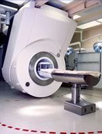
From Philips Medical Systems;
this active shielded member of the Panorama product line combines the advantages of one 1.0 T system's with the possibilities of an open MRI system. The open design helps ease anxiety for claustrophobic patients and increased patient comfort whereby the field strength provides spectacular image quality and fast patient throughput.
Device Information and Specification CLINICAL APPLICATION Whole body Vertically opposed solenoids, head, head-neck, extremity, neck, body/ spine M-XL, shoulder, bilateral breast, wrist, TMJ, flex XS-S-M-L-XL-XXL SE, FE, IR, STIR, FFE, DEFFE, DESE, TSE, DETSE, Single shot SE, DRIVE, Balanced FFE, MRCP, FLAIR, Turbo FLAIR, IR-TSE, T1-STIR TSE, T2-STIR TSE, Diffusion Imaging, 3D SE, 3D FFE, Contrast Perfusion Analysis, MTC;; Angiography: CE-ANGIO, MRA 2D, 3D TOFOpen x 47 cm x infinite (side-first patient entry) POWER REQUIREMENTS 400/480 V | | | |
• View the DATABASE results for 'Panorama 1.0T™' (2).
| | | | |
| | | Searchterm 'Perfusion Imaging' was also found in the following services: | | | | |
| | |
| |
|
| | | | | | | |
• View the DATABASE results for 'Lung Imaging' (7).
| | |
• View the NEWS results for 'Lung Imaging' (3).
| | | | | | Further Reading: | | Basics:
|
|
News & More:
|  |
Chest MRI a viable alternative to chest CT in COVID-19 pneumonia follow-up
Monday, 21 September 2020 by www.healthimaging.com | | |
CT Imaging Features of 2019 Novel Corona virus (2019-nCoV)
Tuesday, 4 February 2020 by pubs.rsna.org | | |
Polarean Imaging Phase III Trial Results Point to Potential Improvements in Lung Imaging
Wednesday, 29 January 2020 by www.diagnosticimaging.com | | |
Low Power MRI Helps Image Lungs, Brings Costs Down
Thursday, 10 October 2019 by www.medgadget.com | | |
Chest MRI Using Multivane-XD, a Novel T2-Weighted Free Breathing MR Sequence
Thursday, 11 July 2019 by www.sciencedirect.co | | |
Researchers Review Importance of Non-Invasive Imaging in Diagnosis and Management of PAH
Wednesday, 11 March 2015 by lungdiseasenews.com | | |
New MRI Approach Reveals Bronchiectasis' Key Features Within the Lung
Thursday, 13 November 2014 by lungdiseasenews.com | | |
MRI techniques improve pulmonary embolism detection
Monday, 19 March 2012 by medicalxpress.com |
|
News & More:
| |
| |
| | | | |
| | |
| | | |
|
| |
 | Look
Ups |
| |